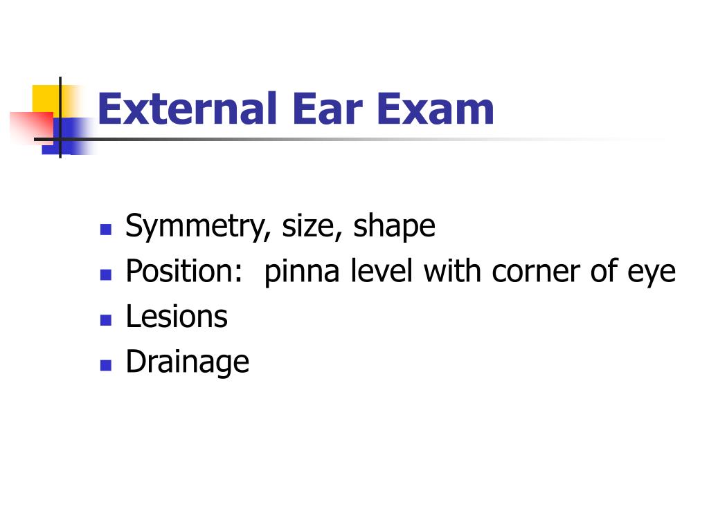
Dispose of any used speculums in the clinical waste bin. Repeat steps 12-18 on the other ear, using a clean speculum. Assess the tympanic membrane for colour changes and shape changes. Identify the pars tensa and pars flaccida. Inspect the walls of the external auditory canal for erythema and oedema.  Inspect the external auditory canal for excess earwax and foreign bodies. Attach a sterile speculum to the otoscope. Optional: Palpate the pre-auricular and post-auricular lymph nodes. Inspect for discharge from the external auditory canals. Inspect both pinnae for colour changes and shape changes. Optional: Test for facial nerve weakness. Check if the patient is in pain or discomfort. It might feel slightly uncomfortable but it should not be painful. Explain procedure and obtain consent: “This will involve me placing a small device inside your ear so that I can see inside it. Can I confirm your name and DOB? Thank you.” I’ve been asked to examine your ears today. Introduction: “Hello, I’m SimpleOSCE and I am a medical student. Grip close to the eyepiece to grant better control. Hold the otoscope horizontally between your thumb, index and middle finger (like you would hold a pencil).
Ask the patient to keep their head as still as possible. Select a sterile speculum which will best fit inside the patient’s external auditory canal.
Ear practice exam skin#
Palpate the pre-auricular and post-auricular lymph nodes for lymphadenopathy (local infection or metastasis of skin cancer).Įnsure this process is carried out on both ears. PalpationĪssess the tragus for tenderness, a classic sign of acute otitis externa.

One of the few painless ear infections, labyrinthitis usually results in vertigo and dizziness. Labyrinthitis (inner ear inflammation): Infection of the labyrinth and vestibular system. Whilst not due to infection, OME can commonly follow acute otitis media. Also known as “glue ear,” OME is a common cause of hearing loss in children.
Otitis media with effusion (OME) is a chronic, middle ear inflammation with an effusion, which leads to a fluid build-up behind the tympanic membrane. The patient will present with a fever, otalgia, conductive hearing loss and a bulging tympanic membrane. The normal functioning of the pharyngotympanic (Eustachian) tube is impaired, increasing the risk of bacterial transit into the middle ear space. Acute otitis media is caused by infection, and is a common complication of a viral respiratory illness. Otitis media (middle ear inflammation): There are many types of otitis media, the most common of which are acute otitis media and otitis media with effusion. Typically, there is tenderness of the tragus and often a conductive hearing loss. Otitis externa (external ear inflammation): Also known as “swimmer’s ear.” This is an inflammation of the external auditory canal, usually due to infection with Pseudomonas aeruginosa or Staphylococcus spp.


 0 kommentar(er)
